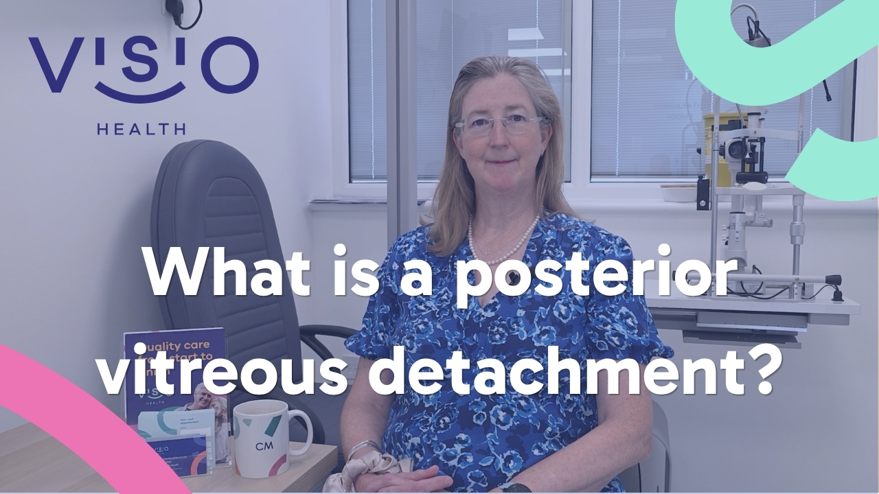
A posterior vitreous detachment, or PVD, happens when the vitreous gel inside the eye changes with age and separates from the retina. It commonly causes new floaters or flashes, but the retina must be checked because a tear can occasionally occur at the same time.
Written by Sanjog Sandhu. Clinically reviewed and approved by Ms Cordelia McKechnie.
Key points
• A posterior vitreous detachment, or PVD, happens when the vitreous gel inside the eye changes with age and separates from the retina.
• Assessment looks at when the flashes or floaters began and whether they are increasing, vision in each eye and whether there is blur, a shadow or a missing area, a dilated examination of the peripheral retina.
• Most posterior vitreous detachments do not need an operation, but the symptoms overlap with retinal tears and detachment.
• First-time or suddenly increasing flashes or floaters, a dark curtain or shadow, sudden blur, eye pain, or symptoms after eye surgery or injury require urgent assessment.
Understanding the question
Floaters and flashes are often caused by age-related change in the vitreous gel, called posterior vitreous detachment or PVD. Most PVDs do not cause a serious problem, but similar symptoms can occur when the retina tears or detaches.
Your symptoms, examination findings and priorities need to be considered together before advice or treatment is recommended.
What the assessment looks for
A new or sudden change needs a prompt dilated retinal examination. The clinician may also use retinal imaging or OCT, depending on the symptoms and findings, but imaging does not replace careful examination of the peripheral retina.
The purpose of testing is not to collect numbers. It is to decide which finding explains the person’s concern, whether another condition is present and which options remain sensible. A result should therefore be discussed alongside symptoms and examination, not read in isolation.
Why the definition changes the next step
Understanding flashes, floaters and the peripheral retina matters because nearby conditions can sound similar but need different decisions. A patient-friendly label is helpful, yet the examination establishes the cause, severity and effect on the rest of the eye. That is what turns a definition into a safe plan.
What may influence the individual plan
The answer can change according to:
• whether the vitreous has separated without damaging the retina;
• whether a retinal tear, bleeding or detachment is present;
• whether observation, laser, freezing treatment or surgery is needed;
• what safety-net symptoms and follow-up apply to that eye.
This is why a general video can explain the principles but cannot replace an examination or a personalised consent discussion.
Treatment, monitoring and next steps
A PVD without a tear is commonly monitored with safety-net advice. A retinal tear may need laser or freezing treatment, while a retinal detachment usually needs urgent surgery. The correct route depends on the examination, not on symptoms alone.
Most posterior vitreous detachments do not need an operation, but the symptoms overlap with retinal tears and detachment. A dilated peripheral retinal examination is what separates reassurance from urgent treatment.
If observation is chosen, the plan should say what is being monitored, when it will be reviewed and which change should bring the appointment forward. If treatment is advised, the patient should understand its purpose, alternatives, likely recovery and the important risks for that eye.
Preparing for a useful consultation
You can make the first appointment more useful if you:
• record whether the change was sudden and which eye is affected;
• do not drive if the vision is obscured or dilating drops are likely;
• bring details of previous retinal or cataract surgery;
• seek urgent care rather than waiting for a routine slot if symptoms are new or worsening.
At VISIO, adults can normally contact the patient-services team directly. An optometrist, GP or another consultant can also refer. If you are using insurance, check referral and pre-authorisation requirements with the insurer before the appointment. Dilation may be needed, so ask whether you should arrange transport.
Questions worth bringing with you
• What is the most likely explanation for my symptoms or test result?
• Which findings matter most, and is anything else contributing?
• What are the reasonable choices, including observation?
• What result is realistic for my eye, and what cannot be promised?
• Which change should make me seek help sooner?
It is reasonable to take notes or bring someone you trust. The goal is not to make a rushed decision. It is to leave knowing what the consultant thinks, why, and what happens next.
When to seek urgent help
First-time or suddenly increasing flashes or floaters, a dark curtain or shadow, sudden blur, eye pain, or symptoms after eye surgery or injury require urgent assessment. Use NHS 111 or an urgent eye service if immediate specialist assessment is not available.
VISIO is not a 24-hour emergency service. If an urgent problem occurs when the clinic cannot assess you promptly, use NHS 111 or the nearest emergency eye service or A&E as appropriate. Call 999 for a life-threatening emergency.
Frequently asked questions
Can this be diagnosed from a description alone?
No. The description can guide urgency, but diagnosis requires the appropriate eye examination and sometimes imaging or measurements.
Can both eyes be affected?
Many eye conditions can affect both eyes but not always to the same degree or at the same time. Each eye is assessed separately.
Are long-standing floaters always an emergency?
Long-standing, unchanged floaters are often less concerning, but any sudden increase, new flashes, blur or shadow needs urgent assessment.
Why are dilating drops used?
They widen the pupil so the clinician can examine more of the peripheral retina. They can blur near vision and increase glare for several hours.
Will an OCT scan rule out a retinal tear?
No. OCT is useful for the macula and other structures, but it does not replace a careful dilated examination of the peripheral retina.
Can a retinal tear be treated before the retina detaches?
Some retinal tears can be sealed with laser or freezing treatment. The correct treatment depends on the tear and the surrounding retina.
Book or ask VISIO Health
If this question is affecting your sight, comfort or confidence, contact VISIO Health in Wooburn Green for advice about the appropriate consultant assessment.
Call or WhatsApp: 0800 032 2020
Email: [email protected]
Online: use the Book a Consultation or Contact form
Sources and editorial method
This guide is based on Ms Cordelia McKechnie’s recorded answer to Q1, expanded for patient clarity using approved VISIO facts and the UK guidance listed below. The expansion explains context, assessment, choices and safety-netting without changing the consultant’s clinical meaning.
• NHS: Floaters and flashes: https://www.nhs.uk/symptoms/floaters-and-flashes-in-the-eyes/
• NHS: Detached retina: https://www.nhs.uk/conditions/detached-retina-retinal-detachment/
• Supporting editorial context: NHS guidance on floaters, flashes and retinal detachment, plus current NHS eye-hospital PVD information.
• Approved VISIO source: Website Content Source Of Truth, checked for the current service pathway and practical details.